
UNFUCG
DashboardSearchChatBookmarksNotificationsActivityPremiumProfile
?
Home
Search
Chat
Saved
Profile
Episode
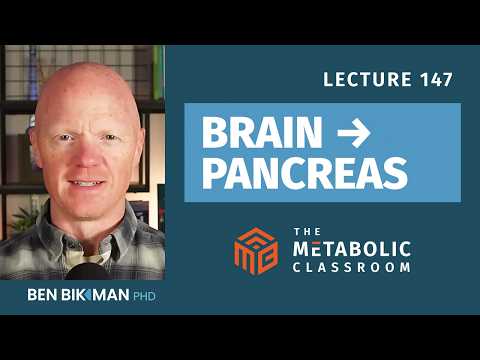
147: How Your Brain Talks to Your Pancreas (The Vagus Nerve Explained)
~26 min
Episode Brief·YouTube

147: How Your Brain Talks to Your Pancreas (The Vagus Nerve Explained)

Ben Bikman
Watch on YouTube Add to chat My bookmarks← All sources

TL;DR

The four things you'd lose by not watching

4 items

TL;DR

The four things you'd lose by not watching

4 items
1

The vagus nerve is the primary communication cable between the brain and metabolic organs, with ~80% of its abdominal fibers carrying sensory information upward, not motor commands down.

2

The cephalic phase insulin response (CPIR) is a small, neurally-driven insulin release triggered by food anticipation, but it's impaired in obesity due to interleukin-1 beta-driven neuroinflammation.

3

Surgically cutting or blocking the vagus (vagotomy, VBLOC) reduces insulin response to oral glucose and can lead to ~25% excess weight loss, proving the vagus is a metabolic lever.

4

Non-invasive vagal stimulation devices like gammaCore (neck) and ear clips are emerging, but metabolic benefits remain preliminary.

Protocols

Concrete recipes — what, when, how much, and why

2 items

Weight Loss to Restore Vagal Tone and Cephalic Phase Insulin Response

WhatLose excess body weight to reduce neuroinflammation and improve vagally-mediated insulin secretion.
WhenFor individuals with obesity or insulin resistance.
DoseNot specified; sustained weight loss likely required.
For whomPeople with obesity, insulin resistance, or type 2 diabetes.
WhyObesity is associated with impaired vagal reflexes and a diminished cephalic phase insulin response due to interleukin-1 beta-driven neuroinflammation. Weight loss can reduce systemic and neural inflammation, potentially restoring normal vagal signaling and improving post-meal glucose control.
CaveatsWeight loss should be achieved through sustainable lifestyle changes; rapid weight loss methods may not address inflammation. Direct evidence in humans is limited, but animal models show reversibility.

Bikman highlights that obesity disrupts the vagus-pancreas axis, with animal studies showing that the vagal reflex to oral glucose is uncoupled and the cephalic phase is attenuated. The defect was linked to interleukin-1 beta in the brain. He suggests that restoring this axis could be achieved through weight loss, which would reduce the chronic low-grade inflammation that impairs neural control. While human data is still emerging, the logic is that weight loss improves heart rate variability (a marker of vagal tone) and glycemic control, consistent with restored vagal function.

Mechanism

Weight loss reduces adipose tissue inflammation and circulating pro-inflammatory cytokines like interleukin-1 beta. This alleviates neuroinflammation in brain regions controlling vagal output, allowing restoration of cholinergic signaling to the pancreas and liver, thereby improving cephalic phase and vagal reflexes.

Restoring it, whether through weight loss or anti-inflammatory strategies or even perhaps direct neuromodulation, that's a topic worth discussing.

Also said
“Lower parasympathetic tone is associated with higher fasting glucose and worse glycemic control. And these associations hold even after adjusting for something like BMI and age.”— Shows that reduced vagal tone correlates with poor metabolic health, independent of obesity, but weight loss could improve it.

Anti-Inflammatory Strategies to Improve Vagal Signaling

WhatAdopt anti-inflammatory lifestyle habits (e.g., diet, exercise, stress reduction) to lower neuroinflammation and enhance vagal control of insulin secretion.
WhenDaily, as part of metabolic health management.
DoseNot specified; chronic inflammation requires sustained intervention.
For whomIndividuals with obesity, metabolic syndrome, or signs of chronic inflammation.
WhyInterleukin-1 beta-driven neuroinflammation impairs the cephalic phase and vagal reflexes. Reducing inflammation could restore these neural pathways, improving postprandial glucose handling.
CaveatsSpecific anti-inflammatory protocols (e.g., drugs like IL-1 beta inhibitors) are not recommended for general use; lifestyle approaches are safer but less targeted. Human evidence is indirect.

Bikman discusses that in animal models, chronic inhibition of interleukin-1 beta normalized the cephalic phase response. While pharmacologic inhibition is not yet a standard human therapy, the principle suggests that any strategy that lowers neuroinflammation could be beneficial. He mentions anti-inflammatory strategies alongside weight loss as a way to restore vagal function. This aligns with broader metabolic health advice, though he doesn't prescribe a specific regimen.

Mechanism

Chronic inflammation, particularly elevated interleukin-1 beta, disrupts neural circuits in the brainstem and hypothalamus that regulate vagal output. By reducing systemic inflammation through diet (e.g., low-glycemic, anti-inflammatory foods), exercise, and stress management, the brain's immune environment may normalize, allowing proper vagal signaling to the pancreas and liver.

Chronic inhibition of interleukin-1 beta normalized that response in that animal model.

Also said
“This is a provocative observation because it connects the chronic low-grade inflammation, even neuroinflammation, which is well documented in obesity, to a specific defect in the neural control of the pancreas.”— Highlights the link between inflammation and vagal dysfunction.

What's new

Personal practice updates, fresh positions, predictions

5 items

Cephalic Phase Insulin Response (CPIR)

A small, early insulin release triggered by food-related stimuli before blood glucose rises, mediated by the vagus nerve; its existence in humans is controversial and it is impaired in obesity.

Why this matters: Challenges the assumption that insulin is only released in response to elevated blood glucose; highlights a neural anticipatory mechanism that may be lost in metabolic disease.

Background

Historically described by Pavlov in dogs, where sham feeding elicited pancreatic secretion that was abolished by vagotomy. In humans, the effect is so small (a few microunits/mL) that it can be masked by normal insulin fluctuations, leading to debate about its reality.

Ben Bikman explains that the CPIR occurs within minutes of seeing, smelling, tasting, or even thinking about food. The signal travels from sensory organs to the brain and then down the vagus to the pancreas, releasing acetylcholine onto beta cells to amplify insulin secretion. However, in humans, the insulin rise is exceptionally small and often indistinguishable from spontaneous oscillations. A 2020 meta-analysis concluded that food-related stimuli do reliably elicit a CPIR, but with substantial heterogeneity depending on the stimulus. This heterogeneity might explain individual differences in responses to sweeteners or food cues. Bikman notes that the CPIR is impaired in obesity, potentially due to dysregulated interleukin-1 beta signaling in the brain's immune cells. Restoring this early insulin pulse, even via a tiny subcutaneous injection, could significantly improve post-meal glucose control. The loss of this neurally-mediated insulin pulse may contribute to postprandial hyperglycemia and delayed insulin peaks in early metabolic dysfunction.

You don't just release insulin when you eat. You may release insulin when you think about food. That signal comes from your brain through the vagus nerve straight to your pancreas.

Also said
“In humans, the cephalic phase insulin response has been a little more controversial. The insulin rise from oral stimulation alone is exceptionally small. We're talking maybe just a few microunits per milliliter, but then the problem is that this can be just washed out with normal spontaneous fluctuations in baseline insulin.”— Explains why the CPIR is hard to detect in humans.
“If the cephalic phase effect in humans is real, and it's small, and it's impaired in obesity, then it may be a meaningful contributor to the postprandial hyperglycemia and the delayed insulin peaks that characterize early metabolic dysfunction.”— Connects CPIR impairment to clinical metabolic dysfunction.
“The cephalic phase is modulated by interleukin-1 beta... and they found that the cephalic phase is impaired in obesity, potentially because of a dysregulated interleukin-1 beta signal.”— Identifies a specific inflammatory mechanism for CPIR loss in obesity.

Beta Cells Use Serotonin for Real-Time Insulin Feedback

Pancreatic beta cells co-release serotonin with insulin, which activates vagal sensory fibers to inform the brain about insulin secretion in real time.

Why this matters: Reveals a direct neural feedback loop that bypasses the slower hormonal route, allowing the brain to monitor insulin output instantly.

Background

Typically, the brain senses insulin only when it reaches the brain via the bloodstream, which takes time. This discovery shows a faster, local neural signal.

Bikman highlights a specific population of vagal sensory fibers that directly innervate the pancreatic islets. Beta cells release serotonin alongside insulin. Serotonin, being a neurotransmitter, can activate these vagal afferent fibers, sending a signal to the brain that indicates how much insulin is being secreted. This provides real-time feedback, allowing the brain to adjust metabolic processes without waiting for insulin to circulate. This mechanism underscores the vagus nerve's role as a bidirectional communication line, not just carrying commands to the pancreas but also listening to it. The implications for metabolic regulation are profound, as disruption of this feedback could contribute to dysregulation in obesity or diabetes.

Beta cells release serotonin along with insulin... These sensory fibers appear to provide a real-time feedback signal to the brain about how much insulin the pancreas is actually secreting.

Also said
“It is so fascinating to me that we typically would think, 'Well, the brain only knows how much insulin's coming out when the insulin makes it to the brain.' And yet, because serotonin is also released when insulin is released, that then affects the vagus, sending a signal directly to the brain, telling the brain how much insulin is coming out.”— Emphasizes the counterintuitive nature of this discovery.

Hepatic Branch of Vagus Mostly Innervates Pancreas and Gut

The hepatic branch of the vagus nerve, despite its name, primarily innervates the proximal small intestine, pylorus, and pancreas, with only a small minority of fibers going to the liver.

Why this matters: Corrects a common anatomical misconception and has implications for interpreting vagal stimulation studies.

Background

Many assume the hepatic branch controls liver function, but its main targets are elsewhere.

Bikman points out that the hepatic branch is misleadingly named. The majority of its fibers actually innervate the proximal small intestine, the pylorus, and the pancreas, with only a very small minority reaching the liver. This is important because when researchers manipulate or discuss the hepatic branch, they must consider its effects on gut and pancreas, not just the liver. This anatomical detail is crucial for understanding experiments like the brain-liver circuit, where severing the hepatic branch disrupted hypothalamic control of liver glucose output—likely because the signal was relayed via pancreatic or intestinal afferents, not direct liver innervation.

The hepatic branch is somewhat misleadingly named. The majority of its fibers actually innervate the proximal small intestine, the pylorus, and the pancreas, which only with only a very small minority actually going to the liver.

Hypothalamic Fat Oxidation Sensing Requires Intact Vagus to Control Liver Glucose Output

Inhibiting fat oxidation in the hypothalamus reduces liver glucose production only if the hepatic branch of the vagus is intact, demonstrating a brain-liver neural circuit.

Why this matters: Provides direct evidence that the brain uses the vagus to regulate liver metabolism based on central nutrient sensing.

Background

The liver's glucose output is known to be regulated by hormones, but this shows a hardwired neural pathway from the brain.

Bikman describes an experiment where researchers inhibited fat oxidation in the hypothalamus of rats. This caused a dramatic reduction in liver gluconeogenesis and glucose output, but only when the hepatic branch of the vagus was intact. Severing that branch abolished the effect, proving that the signal from the hypothalamus to the liver travels via the vagus. This circuit allows the brain to adjust liver glucose production in response to its own nutrient status, independent of blood-borne signals. It highlights the vagus as a conduit for central metabolic control, not just a reflex arc.

When researchers inhibited fat oxidation in the hypothalamus of a rat model... the animals' livers dramatically reduced gluconeogenesis and glucose output, but only if the hepatic branch of the vagus was intact.

Also said
“You sever the hepatic branch and then the signal from the hypothalamus to the liver was lost. This is a clear demonstration that the brain uses the vagus to regulate the liver's glucose output in response to central nutrient sensing.”— Reinforces the necessity of the vagus for this brain-liver communication.

Obesity Impairs Vagally-Mediated Insulin Secretion Through Neuroinflammation

In obesity, the vagal reflex that stimulates insulin release in response to oral glucose is disrupted, partly due to interleukin-1 beta-driven neuroinflammation, and this can be reversed by inhibiting that cytokine.

Why this matters: Links chronic low-grade inflammation to a specific neural defect in insulin secretion, offering a potential therapeutic target.

Background

Insulin resistance is usually attributed to peripheral tissues, but this shows a neural component that fails early.

Bikman explains that in lean animals, glucose arriving at the liver via the portal vein triggers a vagal reflex that stimulates insulin release. In obese rats, this reflex is uncoupled, and they fail to mount a proper insulin response to oral glucose. The cephalic phase is also attenuated. The defect was traced to dysregulated interleukin-1 beta signaling in the brain's resident immune cells (microglia). Chronic inhibition of interleukin-1 beta normalized the response in animal models. This suggests that obesity-induced neuroinflammation directly impairs the vagus-pancreas axis, contributing to postprandial hyperglycemia. Restoring vagal function could be achieved through weight loss, anti-inflammatory strategies, or direct neuromodulation.

In obese rats, this reflex is uncoupled and the animals now fail to mount a proper insulin response to an oral glucose load.

Also said
“The cephalic phase itself is attenuated in obesity. People with obesity tend to have impaired cephalic release... In the mouse model, the defect was traced to that dysregulated interleukin-1 beta signaling.”— Connects human obesity to the same mechanism.
“Chronic inhibition of interleukin-1 beta normalized that response in that animal model. This is a provocative observation because it connects the chronic low-grade inflammation... to a specific defect in the neural control of the pancreas.”— Shows reversibility and therapeutic potential.

Recommendations

Products, supplements, and tools mentioned in the episode

3 items

gammaCore (non-invasive vagus nerve stimulator)

Product

A handheld device applied to the neck that delivers transcutaneous vagus nerve stimulation. FDA-cleared for migraine and cluster headache, not specifically for metabolic conditions.

Bikman describes gammaCore as the most established non-invasive vagal stimulation device. It is applied to the side of the neck where the cervical trunk runs close to the skin. In randomized trials, it reduces pain and attack frequency in headache disorders with a good safety profile. The mechanism involves modulation of central pain pathways, not direct metabolic effects. While not indicated for metabolism, it demonstrates that transcutaneous vagal stimulation can produce reproducible clinical effects, paving the way for potential metabolic applications.

vs alternatives

Compared to implanted VBLOC, gammaCore is non-invasive and can be used as needed, but its effects on weight or glucose are unproven.

The most established device is a handheld unit applied to the neck. It's marketed as gamma core with FDA clearance for several headache indications including things like migraines.

Also said
“In randomized trials, it reduces pain and attack frequency compared with sham or placebo with a very good profile.”— Provides evidence of efficacy for its approved indications.
Find gammaCore

Transcutaneous Auricular Vagus Nerve Stimulation (ear clip devices)

Tool

Devices that stimulate the vagus nerve via the outer ear (auricular branch). Not FDA-approved in the US; studied for depression, anxiety, insomnia, and functional dyspepsia, with preliminary metabolic benefits.

Bikman explains that the vagus has a small sensory branch in the outer ear, making it accessible for non-invasive stimulation. Ear clip electrodes are widely studied but lack FDA approval. Some pilot trials in impaired glucose tolerance have shown improvements in glucose tolerance, and acute shifts in heart rate variability suggest increased parasympathetic tone. However, the evidence for metabolic disease is preliminary and needs larger, controlled studies. These devices are available to consumers but should be approached with caution due to limited regulatory oversight.

vs alternatives

Unlike gammaCore (neck), ear stimulation targets a different branch and may have different effects; both are non-invasive, but ear devices are less established and not FDA-cleared.

The second category is the transcutaneous auricular vagus nerve stimulation and that uses a small clip or electrode on the outer ear. These devices are widely studied, but they're not FDA approved within the United States yet.

Also said
“Pilot trials of the daily ear-based stimulation in impaired glucose tolerance have shown improvements in glucose tolerance, but these need to be replicated with larger better controlled studies.”— Indicates potential metabolic benefit but emphasizes need for more research.
Find Transcutaneous

VBLOC (Vagal Nerve Blockade) implantable device

Product

An FDA-approved implantable device that delivers high-frequency electrical pulses to intermittently block vagal signaling, used for obesity treatment. Leads to ~25% excess weight loss sustained over 2 years.

Bikman describes VBLOC as the only FDA-approved vagal procedure specifically for obesity. Electrodes are placed on the anterior and posterior vagal trunks, and high-frequency pulses silence vagal signaling to the upper abdomen, mimicking a reversible vagotomy. Clinical trials show patients lose about 25% of excess body weight with improvements in glycemic control and cardiometabolic outcomes. The weight loss is modest compared to gastric bypass or GLP-1 agonists, but it provides clean proof that interrupting vagal signaling alone can produce meaningful weight loss without anatomical rearrangement of the gut.

vs alternatives

Compared to bariatric surgery, VBLOC is less invasive and reversible, but weight loss is less dramatic. Compared to GLP-1 drugs, it's a device-based approach with different risk profile.

Implanted electrical leads are placed on the interior and posterior vagal trunks, and they then deliver high-frequency pulses that will intermittently silence vagal signaling... patients have been shown to lose up to about a quarter of their excess body weight, so about 25% of their body fat sustained through 2 years.

Also said
“The magnitude is a little it's modest compared to gastric bypass or to even GLP-1 agonists these days, but mechanistically, it's a very clean proof that if just interrupting the vagus alone is sufficient to produce meaningful weight loss without any of the anatomical rearrangement of the guts.”— Highlights the mechanistic significance of the device.
Find VBLOC

Notable quotes

Lines worth pulling out — contrarian, specific, or perfectly phrased

6 items
You don't just release insulin when you eat. You may release insulin when you think about food. That signal comes from your brain through the vagus nerve straight to your pancreas.
Captures the surprising anticipatory neural control of insulin, a central theme of the lecture.
The vagus is predominantly a sensory nerve... roughly 80% of the fibers in the abdominal vagus are afferent, meaning they are carrying information up to the brain, not commands down to the viscera.
Overturns the common assumption that the vagus is mainly a motor nerve; reframes its primary role as sensory.
The hepatic branch is somewhat misleadingly named. The majority of its fibers actually innervate the proximal small intestine, the pylorus, and the pancreas.
A specific anatomical correction that has implications for interpreting research on vagal signaling.
Beta cells release serotonin along with insulin... These sensory fibers appear to provide a real-time feedback signal to the brain about how much insulin the pancreas is actually secreting.
Reveals a novel neural feedback mechanism that bypasses hormonal delays.
If you could restore an early little bump in insulin because of the cephalic effect... you could significantly improve the glycemic response to a glucose load.
Suggests a therapeutic avenue—restoring the cephalic phase—to improve post-meal glucose control.
The vagus is a genuine lever for body weight and glucose regulation.
Succinctly states the overarching thesis that the vagus is a modifiable target for metabolic health.

Sign in to share feedback

Tell us if this brief hit the mark or missed it — feedback feeds back into the next iteration of the prompt.

Topics covered

vagus-nervecephalic-phase-insulin-responsegut-brain-axisliver-metabolismpancreas-innervationobesity-neuroinflammationvagotomyvagal-nerve-blockadetranscutaneous-vagus-nerve-stimulationheart-rate-variabilityincretinsserotonin-feedbackbrain-liver-circuitinterleukin-1-beta
Free account

Make this library yours

Reading is free for everyone. A free account adds the personal layer: save protocols, follow experts, and see how the other experts weigh in on this same topic.

Create a free accountSign in

Where the experts disagree — weekly

One email a week: the sharpest new disagreements and protocols from the library. No spam, unsubscribe anytime.

Educational summary of the cited expert source — not medical advice. Open the source recording linked above and consult a qualified physician before acting on any protocol.